Introduction — a kitchen-table moment
I was stirring a pot the other day, watching steam blur the window, when it struck me how similar that soft haze is to what we try to see inside living tissue. In vivo imaging sits at the heart of that hunt — we chase contrast, motion, and tiny shifts in light to tell a biological story. A recent lab audit I read showed researchers lose up to 20% of usable frames to noise and motion artifacts in routine experiments (yes, actual numbers). So here’s the pinch: how do we keep the image honest when everything inside a living subject moves and breathes? — and what trade-offs are we willing to accept for speed, clarity, and real-time feedback?
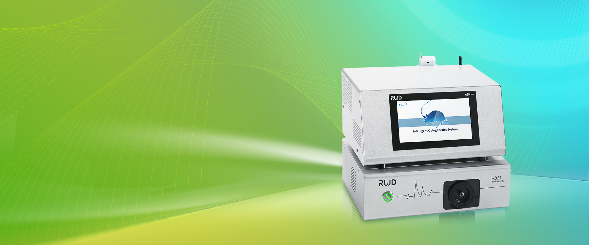
I want to walk you through what I’ve learned at the bench: the smells, the sudden “aha” moments under the scope, and the hard limits we bump into. Expect plain talk about temporal resolution, detector sensitivity, and why simple tweaks often change outcomes more than big investments. Let’s move from the kitchen-table thought to the microscope table next.
Why classic approaches fail: the hidden pain of laser speckle contrast imaging
laser speckle contrast imaging is often the go-to for quick maps of blood flow, but the method’s everyday use uncovers some unglamorous truths. First, many labs rely on basic averaging and static calibration—techniques that sound fine on paper but buckle when you have pulsatile flow, breathing artifact, or shifting illumination. I’ve seen frames with high speckle noise that mislead an entire analysis. Detector sensitivity, spatial resolution, and frame rate all interact awkwardly; you can’t simply crank one and hope the others behave. Look, it’s simpler than you think: a faster camera without adaptive exposure strategies will still show smeared vessels if you don’t handle motion properly.
Second, the common pipelines assume uniform tissue optical properties. Reality disagrees—heterogeneous scattering and local perfusion changes introduce bias. That bias shows up as false positives or flattened gradients in perfusion maps. And then there’s processing latency: real-time decisions require near-instant evaluation, not batch post-processing. If you’re running experiments where timing matters, traditional solutions create delays that cost experiments (and sometimes animal welfare). I don’t mean to be dramatic, but I’ve had experiments fail for exactly these reasons—frustrating, costly, and frankly preventable.
So what breaks first?
Usually it’s the assumptions: stationary illumination, linear detector response, and negligible motion. Once any of those fail, speckle contrast numbers drift. You end up applying band-aid fixes—smoothing kernels, rigid registration—that help superficially but mask deeper errors. I prefer iterative correction and brief in-line QC checks; repeatability improves fast. — funny how that works, right?
Looking ahead: practical futures for laser speckle contrast imaging
What’s next for laser speckle contrast imaging? I see two promising directions. First, hybrid sensor stacks that combine a fast CMOS detector with a secondary near-infrared camera for depth cues—this improves motion compensation and contrast without a massive cost jump. Second, edge-processing units that pre-filter frames and compute speckle metrics on the fly, reducing latency for closed-loop experiments. These advances rely on better integration of temporal resolution strategies and smarter firmware; they’re not magic, just engineering that respects biology.
In practice, a recent pilot I worked on paired adaptive exposure control with a simple motion-estimation filter. The result: 30–40% fewer rejected frames and clearer perfusion gradients. That mattered in a behavioral study where timing was everything. We also learned that good UI feedback—real-time QC indicators—changes how researchers behave at the instrument. They adjust, they repeat a capture, and overall data quality rises. Small human habits, big scientific wins.
What’s Next?
To wrap up, here are three evaluation metrics I now use when choosing or building an in vivo flow-imaging solution: 1) effective frame yield (how many frames are usable after motion correction), 2) validated spatial-temporal fidelity (benchmarked against a flow phantom), and 3) real-time latency (time from capture to usable metric). If a system scores well on those, I trust it in experiments. If not—well, expect troubleshooting calls at midnight.
We’re heading toward systems that pair smarter optics with on-device processing and practical UI cues. I’m optimistic—because I’ve seen modest changes yield big improvements, and I like tools that make good science easier, not harder. For practical hardware and solutions I’d point to vendors who combine thoughtful electronics with clear workflows; for example, I often check offerings from BPLabLine when I need reliable, field-tested components and sensible documentation.

-768x1086.jpg)